
Student preparation
mostafa raeed othman

Student preparation
fatima toameh al toameh

Student preparation
nour kamel deab

Student preparation
fatima omar anan

Student preparation
mohammed belal osama zaror
Radiography is an essential tool in dentistry and a cornerstone of modern dental practices. It enables dentists to view internal structures of teeth, jaws, and surrounding tissues, aiding in the diagnosis of various clinical conditions such as cavities, gum diseases, fractures, and root issues. It is also used in planning treatments like dental implants and orthodontics.
History of Radiography in Dentistry:
- Discovery of X-rays (1895):
In 1895, German physicist Wilhelm Conrad Roentgen discovered X-rays, revolutionizing medicine and dentistry. These rays were first utilized in general medicine, including dentistry, to detect internal diseases and fractures.
- First Dental Radiograph (1896):
In 1896, French dentist Émile G. Dremelin took the first dental radiograph of a patient’s teeth using X-rays, marking the start of specialized dental radiography.
- Conventional Radiography (Film-Based):
For decades, photographic films were used to capture radiographic images. These films required darkroom processing, which was time-consuming and demanded high precision.
- Transition to Digital Imaging:
Advances in technology in recent decades led to the development of digital radiography in dentistry. By the early 1990s, digital devices improved image accuracy and reduced reliance on chemicals used in traditional imaging.
- CBCT (Cone Beam Computed Tomography):
The early 2000s saw the introduction of CBCT technology, offering 3D images for precise diagnostics, especially in dental implants and jaw treatments.
- Immediate Radiography:
Recently, faster, more patient-friendly radiographic methods have emerged, with techniques that expose patients to less radiation than traditional methods.
McDavid, W.D., & Price, J.B. (2013)..
Types of Radiography in Dentistry:
- Intraoral Radiography:
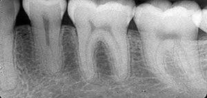
Periapical Radiography:
Examines the entire tooth from crown to root and surrounding tissues, detecting abnormalities in roots or bone structure (Perry, 2020).
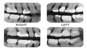
Bitewing Radiography:
Focuses on posterior teeth, identifying cavities and changes in bone thickness related to gum diseases, and determining crown or restoration suitability.
Occlusal Radiography:
Captures the floor or roof of the mouth, detecting cysts or tumors and measuring jaw alignment.
المرجع:
(perry.2020)
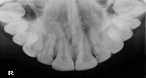
- Extraoral Radiography
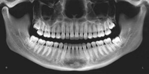
Panoramic Radiography:
Provides a full view of the jaws and teeth in one image, aiding in tumor diagnosis.
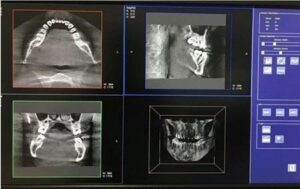
Magnetic Resonance Imaging (MRI):
Produces 3D images of the mouth, jaws, and soft tissues, ideal for implant planning and fracture evaluation (Lannucci & Howerton, 2018).
Uses of Radiography in Dentistry:
- Cavity Detection:
Radiography reveals cavities invisible during visual inspections, especially in interdental spaces or beneath fillings.
- Gum Health Evaluation:
Helps diagnose gum diseases like gingivitis and periodontitis.
- Monitoring Tooth Development:
Tracks tooth growth in children and adolescents to detect developmental issues.
- Implant Planning:
Provides accurate data on bone density and nerve locations for safe and effective dental implants.
- Jaw Disorders Diagnosis:
Detects jaw issues such as temporomandibular joint (TMJ) disorders.
- Orthodontic Treatment Planning:
Assesses teeth and jaw alignment for tailored orthodontic treatments.
- Monitoring Past Treatments:
Ensures the success of previous procedures like root canal treatments or implants.
Tumor and Abnormality Detection:
Identifies tumors or unusual deformities in the oral and jaw regions.
- Locating Impacted Teeth:
Pinpoints the position of impacted teeth, such as wisdom teeth, for potential extraction.
Lannucci and howerton.2018

Risks of Dental Radiography
- Radiation Exposure:
Although generally safe, repeated or unnecessary exposure can increase cancer risks over time.
- Allergic Reactions:
Rarely, patients may react to materials used during imaging, such as in CT scans.
- Misdiagnosis:
Improper interpretation of images can lead to incorrect treatments.
- Anxiety:
Some patients may feel anxious during imaging, especially if they have had negative past experiences.
Pregnancy Concerns:
Pregnant women should avoid radiographs unless absolutely necessary, as radiation exposure may harm the fetus.
Kumar and parasad.2021
Caution should be taken when performing x-rays on children, as their bodies are more sensitive to radiation.
Utilize advanced technologies to minimize radiation dosage:
Conduct radiographs only when necessary.
Use lead shields to protect sensitive body parts.
Discussing potential risks and benefits with a dentist is crucial before undergoing any radiographic procedure (CSPM, 2019).
Conclusion
Radiography is an indispensable diagnostic and therapeutic tool in dentistry. With continuous technological advancements, these techniques are becoming safer, more effective, and more precise, improving healthcare quality while minimizing risks.