Student preparation
Asma Alobide
Student preparation
Miran Aziz
Student preparation
Besan ghnem
Student preparation
Nour Alhuda
Table of Contents:
- General Introduction on the Importance of Medical Imaging in Diagnosis and Treatment
- Its Significance
- Applications
- An Overview of Imaging Technology Development
- CBCT as One of the Latest Techniques
Objective of the Research :
- Highlighting the Benefits of CBCT
- Explaining Its Mechanism
- Its Applications in Dentistry
- Addressing Safety Concerns
Cone Beam Computed Tomography (CBCT):
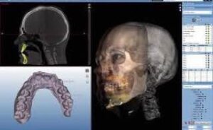
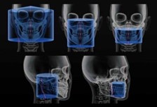
CBCT is an advanced imaging technology used in medicine and dentistry. It provides 3D high-resolution images of internal structures. This technique is primarily utilized in dentistry and oral surgery as a sophisticated alternative to traditional X-rays and computed tomography (CT), offering more clarity and accuracy in diagnosis
CBCT works by emitting a cone-shaped beam around the target area, capturing multiple radiographic images from different angles. These images are then combined to create a 3D visualization that doctors can analyze for various health issues, such as cavities, gum disease, tumors, bone abnormalities, and more.
Significance of Radiographic Imaging in Dentistry:
1-Early Diagnosis: Enables early detection of diseases and conditions, such as cavities, abscesses, and jaw tumors, allowing for cost-effective and efficient treatments.
2-Treatment Planning: Assists in creating accurate and tailored treatment plans for each patient, particularly in surgeries, orthodontics, and dental implant placements.
3-Bone Health Assessment: Critical for evaluating bone density, especially for patients requiring dental implants, ensuring the bone can support the implants.
4-Treatment Monitoring: Allows dentists to track treatment progress, such as the success of root canal therapy or post-surgery healing.
5-Complication Prevention: Identifies hidden problems before they escalate into severe complications, enhancing patient safety and health.
6-Radiographic imaging is an integral part of modern dentistry, providing precise information that improves care quality and minimizes risks.
Medical Applications of CBCT:
1-Dentistry:
Dental Implant Planning: Determines bone quantity and density, nerve locations, and other vital structures.
Assessment of Impacted Teeth: For unerupted teeth like canines or wisdom teeth.
Orthodontic Analysis: Evaluates bones and teeth while monitoring tooth movement during treatment.
Diagnosis of Dental Lesions: Such as cysts, tumors, and bone inflammations.
TMJ Disorders: Detects abnormalities or changes in the temporomandibular
2- Maxillofacial and Oral Surgery:
Fracture Evaluation: Accurately determines fracture location and severity.
Surgical Planning: Enhances outcomes for reconstructive and corrective surgeries.
Tumor Diagnosis: Locates and assesses tumor spread
3- ENT (Ear, Nose, and Throat):
Sinus Evaluation: Diagnoses chronic or acute sinusitis.
Middle Ear Issues: Identifies chronic infections or tumors.
Nasal Septum Deviation: Assessed before surgery.
4- Other Diseases:
Jaw and Bone Disorders: Examines degenerative or congenital conditions.
Infections or Abscesses: Detects infections in bones and tissues

Stages of development of CBCT :
1. CBCT first appeared in the early 1990s as a diagnostic tool to improve 3D imaging.1
The primary goal was to improve imaging in areas such as dentistry, otolaryngology, and maxillofacial surgery.
The initial device was large and uncomfortable, with limited image quality
2.CBCT machines have evolved to become smaller and more efficient
Modern digital sensors have been added instead of traditional films, which has improved accuracy and clarity.
Advances in software have reduced imaging time and made it possible to reconstruct images with high accuracy
3.The use has expanded to include maxillofacial surgery, surgical planning, and upper respiratory imaging.
The technology helps doctors make accurate treatment decisions based on 3D data.
4.Optimize radiation dose
One of the major issues faced with CBCT is the high dose of radiation compared to conventional X-rays.
Technology has evolved to reduce radiation dose without compromising image quality.Low dose imaging modes have been added to accommodate pediatric and pregnant patients.
5.The future
CBCT is expected to develop into more specialized and accurate, with techniques allowing imaging of soft tissues.
Integration with virtual and augmented reality (VR/AR) technology to provide more accurate surgical planning.
Reduce the size of devices and make them more portable and usable in multiple places
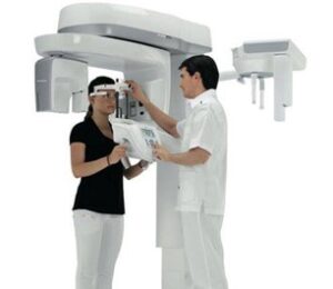
Steps to perform CBCT:
1-Patient preparation The doctor asks the patient to remove any metal objects such as glasses or earrings.
2-Patient position The patient sits or stands in the device with the head fixed to ensure no movement.
3-Imaging process The device rotates around the patient’s head to collect data within seconds
4-Data processing The data is sent to the computer to collect images and display them in 3D.